INSTRUCTIONS AND PROPHECIES OF THE Blessed MOTHER ALIPIA GOLOSEEVSKY, Kyiv...


Content
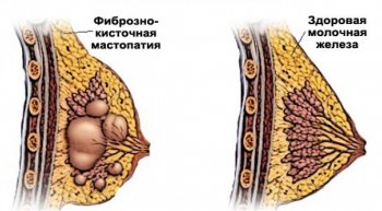
Fibrocystic mastopathy (FCM) is a benign disease, the main symptom of which is pathological changes in breast tissue in the form of violations of the ratio of connective tissue and epithelial components. This disease can occur in women of any age - both in adolescents and during menopause. Progesterone deficiency, increased production of estrogen, androgen can provoke a disease, about treatment, diagnostic methods, the types of which you will learn from the article.
The following signs will help to suspect the presence of FCM, including bilateral, which can occur both individually and all together:
The diagnosis can only be made by a qualified doctor. However, the first harbingers in the form of increasing constant pain, the condition of the chest will let the woman know that something is not happening properly. Diagnosis includes the following methods: palpation, primary examination, ultrasound diagnosis of glandular tissue, mammography. Pay attention to the appearance of the breast, signs of its enlargement, asymmetry, the condition and location of the nipples, the color of the halo, the presence of discharge.
At the first concern in the chest area, every woman is obliged to consult a doctor in order to begin treatment of the disease at an early stage. To make a diagnosis based on the patient's complaints, an additional examination is prescribed:
Due to the similarity of breast cancer and nodular mastopathy, it is necessary to conduct a thorough examination by a mammologist using radiological, clinical, morphological, cytological, echographic methods. In the case of nodular mastopathy, foci of compaction are determined, which have clear boundaries, a lobed, smooth or granular surface. With an intraductal location, pressure on the peripapillary zone is accompanied by secretions of different colors and textures.
During the survey mammography, zones of intense homogeneous darkening, calcifications, oval shadows of cysts, fibrous bands can be found on radiographs. The combination of different forms of the disease gives a vivid picture, which is characterized by multiple darkened areas, restructuring of the structure of the gland, areas of enlightenment of various shapes, sizes, the presence of separate shadows of fibroadenomas, strands of connective tissues, cysts.
If a cyst is detected, a puncture with a cytological examination is performed, after which a pneumocystography is performed. The latter is necessary to control the completeness of the emptying of the formation, the detection of tumors, intracystic hyperplastic formations. If intraductal changes are suspected, ductography is performed. It is determined by the introduction of contrast into the ducts, their expansion, deformation, cystic cavities, deposition of calcium salts. Doppler ultrasound of the glands makes it possible to judge the size, location, vascularization of formations, their structure.
ICD-10 is the tenth revision of the international classification of the disease (according to Wikipedia). In Russia, it is adopted as a single regulatory document in order to take into account the reasons for the appeal of the population, death, and morbidity. According to the classification, mastopathy is included in the section Benign breast dysplasia N60:

The main reason for the development of FCM lies in changes in the hormonal background, a reflection of which is the menstrual cycle. Such hormonal variability is primarily manifested through a violation of the ovarian-menstrual cycle, which is eliminated on its own. Disturbances in the work of hormones can manifest themselves under the guise of premenstrual syndrome, which causes a lot of difficulties and worries for a woman. Fibrocystic mastopathy means that periodic disturbances have become permanent, making the breast a vulnerable, weak link.
The cause of the disease may lie in the state of the spine, since the regulation of metabolic processes in the chest comes from the thoracic vertebral region. The next factor that can provoke the disease is a violation of the water balance. If a woman does not have the habit of drinking water regularly, then the body is in a state of water starvation, and this leads to changes in hormonal levels. Mastopathy can be provoked by chronic stress, endocrine diseases, problems of the reproductive organs, endometriosis.
Thanks to many years of experience in studying this disease, mammologists have at their disposal several methods of treating FCM. To determine a specific program to get rid of the pathology, a complete examination should be carried out, the cause of the hormonal imbalance should be eliminated, and the nervous system should be restored. In the treatment of diffuse mastopathy, large dosages of vitamins are recommended to activate the immune system, homeopathic medicines to normalize liver function.

Drug treatment includes hormonal and non-hormonal. The first type is necessary in order to regulate the cyclicity in the system, including the hypothalamus, pituitary gland, ovaries, since this normalizes the hormonal background by influencing the gland tissue. The second type of treatment stops the manifestations of FCM in its mild form.
Non-hormonal conservative therapy includes the following means:

If multiple seals appear in the chest, easily palpable in the outer sections, in the center, this indicates the presence of diffuse FCM. At the initial stage, these changes are unstable, mild, but if left untreated, the seals become rough. With diffuse mastopathy, the use of folk remedies is allowed. The following recipes may help:

Hormonal treatment of FCM is aimed at stabilizing breast tissue and is used after examining the status of hormones. The following drugs are used:

Physiotherapy procedures for patients undergoing FKM treatment are prescribed very rarely. This is due to the fact that this disease is considered a contraindication to thermal manipulations. An exception to the rule are methods of treatment with mud baths, electrophoresis, shock wave therapy, ultrasound therapy, and other procedures.

Surgical treatment of FCM is indicated for the nodular form of the disease, which is practically not amenable to drug therapy. Indications for surgery are those cases when there is a rapid growth of neoplasms, the presence of severe concomitant diseases that do not allow long-term drug treatment, the presence of atypical cells in biopsy samples, the ineffectiveness of other methods, and a high probability of transformation into a cancerous tumor.
In most cases, a sectoral resection is performed. Such an operation involves the removal of a sector of the gland that is affected by nodes and cysts. The surgery takes about 40 minutes and is performed under general anesthesia. As necessary, after the operation, drug therapy is prescribed: antibacterial drugs are prescribed, additionally - medicines to stimulate the immune system, normalize the blood count, complexes of vitamins and minerals.

Special nutrition in the treatment of mastopathy involves the use of certain healthy foods and the restriction of harmful ones. In addition to the contents of the daily menu, you should follow the correct regimen: eat at least five times a day at the same time. During the meal, do not be nervous, move away from irritants, learn to relax. The following products have a positive effect on the general condition, slow down the growth of connective tissues:
To reduce the risk of developing cysts, it is necessary to completely abandon foods that cause an increase in the secretion of female sex hormones: semolina, premium flour products, conservation, pickles, corn oil, confectionery, margarine, mayonnaise, soda, smoked meats, fatty foods, black tea, coffee, white cabbage.
Learn more about what to do with such a diagnosis.
As a rule, mastopathy is not prone to complications. Proliferative and nodular forms of the disease can become malignant over time, turning into breast cancer. However, with proper and timely treatment, the prognosis is favorable. FCM therapy involves lifestyle changes that include the following contraindications:
- pathological fibrocystic changes in the breast tissue, characterized by the appearance of dense, often painful, fine-grained formations. Concerned about engorgement, soreness of the gland, more pronounced before menstruation, serous, sometimes bloody discharge from the nipple. Has a tendency to relapse, is a cancer risk factor. Diagnosis of mastopathy requires mammography, ultrasound of the mammary glands, if necessary - diaphanoscopy, MRI of the mammary glands, pneumocystography, puncture biopsy. Treatment of mastopathy is carried out by conservative methods. If there is a danger of malignancy of nodular mastopathy, the node is surgically removed.

- a concept that combines a group of diseases of the mammary glands, characterized by the development of pathological changes in the gland tissue with a violation of the ratio of epithelial and connective tissue components. According to the WHO nosological classification of 1984, mastopathy is understood as fibrocystic disease of the mammary glands. The incidence of mastopathy of various etiologies in young women ranges from 30-45% and increases markedly after 40-45 years.
Mastopathy is a benign change in the tissue of the gland, which is directly dependent on neurohumoral regulation. This means that the factors in the development of mastopathy are both pathologies associated with disorders of the state of nervous regulation (stress, neurosis, depression), and a disorder in the hormonal balance and internal homeostasis of the body.
There is currently no complete understanding of the causes and mechanisms of development of mastopathy, but there is every reason to believe that hormonal status plays a significant role in the occurrence of this disease. Factors contributing to the development of mastopathy: early menopause, menstrual disorders (hormonal dysfunctions, polycystic ovary syndrome, improper use of hormonal contraceptives), prolonged absence of childbirth, numerous (more than three) abortions, irregular sex life (or its absence), diseases of the genital organs , lactation for less than three months, endocrine pathologies (hypo - and hyperthyroidism, dysfunction of the hypothalamic and pituitary regulation, the work of the adrenal glands, liver, pancreas), hereditary predisposition.
There is an assumption that the most significant pathogenetic factor in the development of mastopathy is progesterone deficiency with an excess of estrogens. In this case, there is an increase in the proliferation (reproduction) of epithelial cells and connective tissue cellular elements. In addition, the production of prolactin plays a significant role in the pathogenesis of mastopathy. Prolactinemia increases the sensitivity of breast tissue to estrogen.
The most common classification of mastopathy in clinical practice distinguishes three forms: mastalgia (mastoplasia or mastodynia), diffuse fibroadenomatosis and localized fibroadenomatosis. Mastalgia is characterized by the predominance of a pronounced pain syndrome and is an indication for the appointment of analgesics.
 Diffuse adenomatosis is the development of diffuse seals and cysts in the gland tissue. It is divided into two types: fibrous mastopathy, when connective tissue seals are predominantly formed in the gland tissue, and fibrocystic mastopathy, if cysts (fluid-filled cavities) form in the gland in addition to foci of fibrosis.
With localized fibroadenomatosis, pathological changes are detected in a limited area of \u200b\u200bthe gland (segment, quadrant) and do not spread throughout the parenchyma of the organ. The detection of a localized mass in the breast is an indication for a biopsy to rule out a malignant tumor.
 The most characteristic symptom of mastopathy is the detection of compaction in the mammary gland during palpation. This hardening can often be painful, and the pain usually gets worse during the second phase of the menstrual cycle and just before a period. The compaction can be single, several nodules can be detected, the entire gland can be felt compacted. Mastopathy is characterized by damage to both glands, mainly their upper sections.
The predominance of the fibrous component is detected by touch as a seal, cystic changes in the early stages may not be detected at all on palpation (microcysts of the ducts). Pain in the mammary glands, as a rule, has a dull, aching or pulling character. Its occurrence is associated with compression of nerve endings in the glandular tissue by fibrous growths, as well as their partial sclerosis. The intensity of the pain syndrome depends on the severity of the pathology, most often the occurrence and intensification of pain is associated with the menstrual cycle (before menstruation at the peak of estrogen production, the pain intensifies). Sometimes there is irradiation of pain in the shoulder blade, arm.
In 10-15% of women, there are no complaints of soreness, although pathological changes of a significant degree are found on examination. This is associated with a different level of pain sensitivity in women and individual branching of the nervous system of the mammary glands. About 10% of mastopathy are accompanied by an increase in lymph nodes in the armpits. Sometimes palpation of the lymph nodes is moderately painful.
An increase in the volume of the mammary gland, their periodic engorgement (in the second period of the menstrual cycle) is associated with the formation of venous congestion in the vascular network of the glands and swelling of the connective tissue. Glands can increase by 15%. This is characterized by a feeling of discomfort and pain on palpation (increased sensitivity of the chest). The combination of these symptoms is called premenstrual syndrome.
You also need to carefully consider the detection of a node (or several). Palpation of a dense limited nodular formation may be a sign of localized nodular mastopathy, and may be developing breast cancer. When identifying nodes suspicious from the point of view of malignancy in the mammary gland, their biopsy is always prescribed.
One of the most significant elements of the timely detection of pathologies and neoplasms in the mammary glands is self-examination (self-palpation of the mammary glands). To identify formations, determine their shape, size, quantity, as well as to identify diffuse pathological changes in the gland tissue, instrumental diagnostic methods are used.
 Biocontrast mammography is an X-ray examination of the mammary glands. Mammography is best done in the first phase of the menstrual cycle. A picture of the chest is taken in two projections: frontal and lateral. This study is one of the most informative and specific.
In addition, ultrasound of the mammary glands is currently used. As a rule, fibrocystic changes in the tissue of the glands affect the echogenicity of its structures and can be detected and studied quite qualitatively using this technique.
MRI of the breast marks areas of increase and decrease in the temperature of the gland tissues. The technique of diaphanoscopy consists in transillumination of the mammary gland using a light source. In this case, the neoplasm in its thickness will be noted as a darker spot. With the help of ductography, the system of the milk ducts is examined. A contrast agent is injected into the mammary gland through the nipple, after which an x-ray is taken. The picture shows the ductal system, areas of deficiency of filling with a contrast agent may be signs of neoplasms. Pneumocystography is performed under ultrasound guidance. Air is introduced into the cyst cavity using a thin needle, which allows you to straighten the walls and carefully examine them for parietal formations.
When a nodular formation is detected, a biopsy of the mammary gland is performed - extraction by puncturing a tissue sample with a thin needle for histological examination. To identify the etiological factors of mastopathy, methods for studying the hormonal status are used. Colposcopy and cytological examination of the cells of the vaginal epithelium allows us to conclude about the total hormonal background, since the shape and structure of the cells directly depend on the action of sex hormones.
They directly determine the content of hormones in the blood: progesterone and estrogen, follicle-stimulating, luteinizing hormones, as well as thyroid hormones and thyroid-stimulating hormone, adrenal hormones. Sometimes a test is performed for the presence of autoantibodies to thyroid cells to detect autoimmune thyroiditis. These specialists jointly produce a thorough analysis of the endocrine system and prescribe drugs that correspond to the identified pathologies.
With severe estrogen (and significant pain), drugs can be prescribed that reduce the effect of these hormones on the mammary gland (tamoxifen, toremifene citrate). To normalize the menstrual cycle, oral contraceptives are used (selected in accordance with the hormonal status). For the treatment of functional disorders of the thyroid gland, drugs that regulate the production of thyrohormones are used. Vitamin complexes help to improve liver function and normalize metabolic processes.
Among other things, topical progesterone preparations are used (they act directly on the gland tissue, helping to reduce the proliferation of connective tissue and epithelial cells, removing swelling), homeopathic remedies. Patients suffering from mastopathy are advised to limit the use of coffee and strong tea, stop smoking, enrich the diet with fruits, vegetables, foods high in fiber and vitamins. If a malignant tumor is suspected, surgical removal of the node is performed, in other cases they are limited to conservative treatment.
As a rule, mastopathy is not prone to complications and malignancy. With proper correction of the hormonal state, the prognosis is positive, but hormonal disruptions can provoke relapses.
Many factors contributing to the development of mastopathy make it difficult to develop a unified and consistent prevention scheme. However, the most significant factors should be avoided: stressful situations (as a preventive measure, it is recommended to take therapeutic sedatives of natural origin - valerian, motherwort), creating a psychologically comfortable environment, a positive way of thinking.
Proper balanced nutrition without excess calories, prevention of excess weight and obesity, however, without being carried away by mono-diets and dubious methods of losing weight, help maintain internal homeostasis and the proper functioning of the neurohumoral regulatory system. One of the components of the diet that negatively affects the hormonal status of women is caffeine. Women should limit, if possible, completely eliminate caffeine from their diet and in no case abuse strong coffee on an empty stomach.
Older women using oral contraceptives should stop smoking. Also useful in terms of preventing breast pathologies will be limiting the use of alcoholic beverages. A significant factor in maintaining a woman's health is regular sexual activity and physical activity.
What is the danger of mastopathy, if it is not treated, are interested in women who first encountered this diagnosis. Mastopathy is a benign formation in the mammary gland, which can cause various complications in the absence of competent therapy.
Mastopathy is characterized by a pathological proliferation of connective and glandular tissue in the chest, the formation of cysts, cavities, seals, and nodules in them.
Allocate diffuse and forms of the disease, the first one is, which is the most common.
The main reason for the appearance of mastopathy is a violation of the normal hormonal balance in the body. Factors that can provoke the development of the disease:
It is impossible to name the exact reasons that caused mastopathy. Diagnosis should be done by a doctor, who must be contacted without delay, when the first signs of the disease appear.
The following symptoms may indicate the development of fibrocystic mastopathy of the mammary glands in a woman:
What will happen if mastopathy is not treated, what are the consequences and prognosis for the patient? Mastopathy is a benign tumor. In the initial stages, it responds well to medical treatment. However, the lack of competent therapy significantly increases the risk of the transition of the neoplasm to a malignant form.
So is mastopathy dangerous or not, and what does this disease lead to? Contacting a mammologist at the first signs of pathology will stop the progression of mastopathy, eliminate symptoms, and improve well-being.
Thus, ignoring the diagnosis of mastopathy leads to a significant increase in the risk of breast cancer. This can be avoided by visiting the doctor in a timely manner and undergoing a breast examination.

 The opinion that mastopathy affects only the mammary glands is erroneous.
The presence of pathological foci has a negative effect on the entire body and causes certain symptoms:
is the only sure way to solve the problem. It is recommended to consult a doctor as soon as possible if there is a suspicion of the development of the disease.
At the initial stages, drug treatment is prescribed, which includes the necessary drugs, as well as vitamins. The patient is given recommendations regarding the diet, the selection of the right underwear.
With advanced forms of mastopathy, it is possible to prescribe surgical intervention.
On the video about the danger of mastopathy
It is much easier to prevent a disease than to treat it later. This rule is also relevant in relation to mastopathy.
Preventive measures include regular examination of the mammary glands. For women over the age of 35, it is necessary to undergo a mammogram once every 2 years, after 50 years - this should be done annually.
What is the danger of mastopathy if it is not treated? Refusal of therapy leads to the possibility of developing oncology. The risk is quite high and is about 60%. Only a timely visit to the doctor, treatment and compliance with the recommendations of the doctor will allow you to get rid of the disease and return to a healthy and fulfilling life.
Mastopathy is a concept that combines a number of certain diseases of the mammary glands, which are characterized by the development of pathological changes in the gland tissue itself. At the same time, the ratio of the components of connective and epithelial tissues is also disturbed. The nosological classification of the World Health Organization characterizes mastopathy as a fibrocystic disease of the mammary glands.
The chance of being affected by various mastopathy of any etiology in a female at a young age fluctuates somewhere within forty percent, but increases significantly after forty years.
Mastopathy in itself is a change in the gland tissue of a benign nature, which is dependent on nervous and humoral regulation. It can be argued that the main factors in the development of mastopathy are pathological processes that are closely related to disorders of nervous regulation (neurosis, stress, depression) and imbalance of hormones (including internal homeostasis).
The most common classification of mastopathy in clinical practice, where there are 3 forms: mastalgia (mastoplasia, mastodynia), as well as diffuse and localized fibroadenomatosis.
Mastalgia is characterized by the predominance of a pronounced pain syndrome, which is the main indication for prescribing special analgesic drugs to the patient.
Diffuse adenomatosis is a process of development of cysts and diffuse seals in the glandular tissues. There are two types: this is fibrous mastopathy (in this case, mainly seals from the connective tissue are formed in the breast tissues) and fibrocystic mastopathy. In the case of the latter, in addition to the foci of fibrosis, cysts (cavities that are filled with fluid) also appear in the mammary gland.
In the case of localized fibroadenomatosis, pathological changes appear only in a very limited area of \u200b\u200bthe mammary gland (quadrant or segment) and do not spread throughout the parenchyma of the gland.
If a localized neoplasm is found in the breast tissue, a biopsy should be performed to exclude the presence of a malignant tumor.
Unfortunately, there is no complete and detailed understanding of the causes and mechanisms of development of mastopathy, but there is every reason to assert the fact that the hormonal status plays a key role in the development of this pathology.
There is an assumption that the most significant pathogenetic cause of the development of mastopathy is a deficiency of the hormone progesterone with an excess of the hormone estrogen. In this case, there is such a phenomenon as increased proliferation (reproduction) of the cellular elements of the connective tissue, as well as epithelial cells. In addition to this phenomenon, the production of prolactin by the body plays an important role in the pathogenesis of mastopathy. Prolactinemia affects the increase in the sensitivity of breast tissue in relation to the hormone estrogen.
The most characteristic symptom of mastopathy is the detection of a compacted area in the tissues of the mammary gland during palpation. Most often, this seal is slightly painful. The pain tends to increase in the second phase of the menstrual cycle, as well as immediately before the menstruation itself. These seals are both single and multiple. Several nodules can be detected, and the entire mammary gland can be palpated compacted. For mastopathy, it is also common for the disease to affect both glands at once, most often their upper sections.
The excessive presence of the fibrous component appears to the touch as a kind of thickening, and changes in the cystic nature in the tissues in the early stages of the disease may not be felt at all during palpation of the gland (microcysts of the ducts).
The pain in the mammary glands itself most often has a aching, dull or even pulling character. The occurrence of pain is associated with compression of nerve endings by fibrous growths in the parenchyma of the mammary gland or even its partial sclerosis. The degree of intensity of the pain syndrome strongly depends on the severity of the pathology itself. Most often, the manifestation and intensification of pain is directly related to the menstrual cycle (before menstruation, pain increases at the peak of estrogen hormone production). Sometimes there is a phenomenon of irradiation of pain in the region of the scapula or hands.
In some examined women, the soreness of the seals is not observed, although when examined by a doctor, pathological changes of varying severity are found. This phenomenon is associated with a distinctive threshold of pain sensitivity, as well as with an individual feature of the branching of the nervous system in the tissues of the mammary glands.
About 10% of mastopathy occur with an increase in lymph nodes in the armpits. Palpation of the lymph nodes is usually moderately painful.
An increase in the size of the mammary glands, their periodic coarsening (usually in the second period of the menstrual cycle) is a consequence of the presence of venous congestion in the vessels of the mammary glands, including edema of the connective tissue. In this case, the mammary glands can increase in size by more than 15%. This is characterized by a feeling of discomfort and even pain during palpation (increased sensitivity of the entire chest). The combination of all these signs will be premenstrual syndrome.
Sometimes there may be discharge from the nipples. They are of various nature, etiology and any degree of abundance. They can appear both when exposed to the nipple itself, and can be quite pronounced. In its consistency, the discharge is usually whitish or completely transparent, but in some cases it can become bloody, brown or greenish in color. The greatest danger is bloody discharge, for the reason that they can be the main sign of the development of malignant processes in the mammary glands. The appearance of absolutely any discharge from the nipples, of any nature, is an important reason for contacting a mammologist.
It is also extremely important to pay attention to the detection of one or more nodes. Palpation of a dense, limited in size, nodular formation can become both a sign of localized nodular mastopathy, and it can turn out to be cancer. If suspicious nodes are found in the tissues of the mammary gland, they are always biopsied in order to exclude malignant tumors.
The main, most significant and main way of timely detection of neoplasms and pathologies in the mammary glands is the method of self-examination (in other words, self-checking (palpation) of the mammary glands).
To detect formations, as well as determine their size, shape, quantity; to find diffuse pathological changes in the parenchymal tissues of the mammary gland, instrumental diagnostic methods are used.
Biocontrast mammography is an examination of the mammary glands using x-rays. Mammography is best done during the first phase of your menstrual cycle. Pictures of each mammary gland are taken in the frontal and lateral projections. This survey is one of the most specific and informative.
In addition to this method, ultrasound of the mammary glands is currently used. Fibrocystic pathologies of breast tissues affect the echogenicity of their structures. For this reason, changes can be detected in time and studied quite qualitatively, thanks to this technique.
MRI of the breast determines the zones of decrease and increase in the temperature of breast tissue.
The method of diaphanoscopy consists in the lumen of the chest using a bright light source. When using this method, the neoplasm in the thickness of the gland will look like a darker spot in color.
With the help of the method of ductography, a study of the system of the lactiferous ducts of the gland is carried out. In this case, contrast is injected into the mammary gland through the nipple, after which an x-ray is taken. The picture shows the milk duct system. Areas in which the contrast agent is not sufficiently expressed may be signs of formations and various pathologies.
Pneumocystography is performed under ultrasound guidance. With this method, air is pumped into the cavity of the cystic formation with a needle. This allows you to sufficiently straighten the walls and examine them well in order to detect parietal neoplasms.
When a nodule is found, a breast biopsy is performed. In this method, using puncture with a fine needle, a sample of gland tissue is removed for histological examination.
When determining the etiological factors in the development of mastopathy, methods for studying the hormonal status of the body are actively used.
Methods of colposcopy and cytological study of the cells of the epithelium of the vagina allow us to make an objective conclusion about the total and general hormonal background, since the shape, size and structure of the cells of the vagina are directly dependent on the influence of sex hormones. This method also directly determines the amount of such hormones in the blood as: estrogen and progesterone, luteinizing, follicle-stimulating hormones, adrenal hormones, as well as thyroid hormones and thyroid-stimulating hormone.
Sometimes a test is done for the presence of autoantibodies to thyroid cells. This is necessary to identify a pathology such as autoimmune thyroiditis.
To find out the general hormonal state of the patient's body, comprehensive studies of the organs of the endocrine system are carried out to identify their possible pathologies. These include: ultrasound of the thyroid gland, liver, adrenal glands; CT scan of the pituitary gland, radiography of the Turkish saddle.
To exclude immune and metabolic pathologies, a biochemical blood test is performed and an immunogram is made.
In the treatment of mastopathy, one of the most important roles is played by the correction of the balance of hormones in the body. When choosing methods of treatment, it is necessary to consult such specialists as a gynecologist and an endocrinologist. Doctors jointly conduct a thorough analysis of the state of the endocrine system, if necessary, prescribe drugs that correspond to the identified pathologies of organs and systems.
With a pronounced form of estrogen, as well as with severe pain, drugs may be prescribed (for example, tamoxifen, faresterone), which reduce the effect of these hormones on the mammary gland.
To normalize the cycle of menstruation, oral contraceptives are used, which are selected depending on the hormonal status of the patient.
For the treatment of thyroid disorders, drugs that regulate the production of thyroid hormones are used.
Vitamin complexes improve liver function, as well as normalize metabolic processes in the body.
Among other methods, homeopathic remedies and local progesterone hormone preparations are used. They act directly on the breast tissue, which helps to reduce the proliferation of epithelial cells and connective tissue, removing puffiness.
Patients who suffer from mastopathy should significantly limit the use of drinks and drugs containing caffeine, as well as stop smoking. It is recommended to enrich the diet with vegetables, fruits and foods that contain a large amount of fiber and vitamins.
If there is a suspicion of a malignant formation, the node is surgically removed. In other cases, treatment is limited to conservative methods.
A large number of factors that contribute to the development of mastopathy complicates the development of a consistent and unified prevention scheme.
Nevertheless, it is easy to single out the most significant factors influencing the prevention of mastopathy: stressful situations (in case of stress, as a preventive measure, it is recommended to use soothing medicinal preparations of biological origin - valerian root, motherwort), a positive way of thinking, psychologically comfortable environment.
A balanced, non-caloric, proper nutrition (but without a passion for a variety of mono-diets and low-quality methods of losing weight), as well as the prevention of obesity and excess weight, support the internal homeostasis of the body and the proper functioning of the regulatory neurohumoral system.
One of the main components of the diet, which negatively affects the hormonal background of patients, is caffeine. Women need to limit or even eliminate caffeine-containing foods from their diet, and should not abuse strong coffee or tea on an empty stomach.
Women who use oral contraceptives should stop smoking. It will also help to limit the consumption of alcoholic beverages.
A very significant factor for maintaining the health of the patient is a regular high-quality sex life and, in general, any long-term physical activity of the body.
Most often, cases of mastopathy do not carry complications and malignancy. The prognosis of the disease is positive, but only with proper management of the hormonal state. However, one should be wary of hormonal disruptions that can provoke relapses.
According to WHO statistics, more than 40% of women suffer from mastopathy, while the risk of its occurrence in patients suffering from various gynecological diseases is much higher - about 60%. Absolutely all women aged 14 to 60 belong to the risk group. The peak incidence occurs in the childbearing age (20-45 years), after which it begins to subside.
The smallest functional unit of the breast is the alveolus. This is a small vesicle, which is lined from the inside with a glandular tissue dotted with lactocytes. In turn, the alveolus consists of myoepithelial cells that have the ability to contract and increase, and thus regulate the volume of the alveoli. In each of them there is a duct, through which myoepithelial cells from time to time throw out the secret that accumulates in them.
The ducts are lined with epithelium, the structure of which is similar to glandular cells. It is here that lactation processes take place.
The connection of 150-200 alveoli forms a lobule, from 30-80 lobules the lobe of the mammary gland is formed, in the middle of which there is a large duct, which is formed from smaller alveolar ducts. Each breast is a formation of 15-20 lobes with milk ducts leading into the nipple.
In addition, in the female breast there is adipose, connective and glandular tissues, the ratio between which is laid down at the genetic level (the main determining factor is the state of the reproductive system). It is they who determine the shape and size of the breast. Violation of the ratio inherent in nature between the connective and epithelial tissues of the mammary gland causes mastopathy. In this article, we will look at the symptoms and treatment of mastopathy, talk about how to avoid complications, what preventive measures will help to avoid the development of the disease.
First phase observed in patients under 30 years of age. They, as a rule, have a regular or almost regular menstrual cycle, to which the main manifestations of the symptoms of the disease are attached.
Second phase observed in women 30-40 years old: at this age, mastopathy manifests itself most clearly.
The main reason that negatively affects the condition of the tissues that make up the mammary gland is an imbalance between the production of progesterone and estrogen. In the first case, there is a decrease in the amount of the hormone, in the second - its significant increase. Often, the hormone prolactin, which directly affects the functional state of the female breast, is also included in the processes. An increase in its amount contributes to engorgement and swelling of the mammary glands, as well as the appearance of pain in them. Pregnancy can cause hormonal failure, which is why mastopathy often manifests itself during this period.
Third phase mastopathy is observed in women over 40 years of age: pain decreases, in the tissues of the mammary gland there are many small cysts filled with a brownish secret.
The age differentiation of the phases of mastopathy is conditional, since some negative factors (poor environmental conditions, abortions, etc.) contribute to the "rejuvenation" of the disease.

Trauma can provoke the appearance of mastopathy - upon impact, a hematoma occurs, which is subsequently involved in the pathological process of growth of the soft tissues of the mammary gland. This includes wearing tight bras.
By the nature of neoplasms and the intensity of growth of new cells, proliferative and non-proliferative forms of mastopathy are distinguished. In the first case, the amount of dense connective tissue increases rather slowly, and in the second - much more intensively. In this regard, non-proliferative mastopathy often transforms into malignant neoplasms, and therefore requires more careful medical supervision.
Fibrous mastopathy- a benign disease of the mammary gland, in which the glandular tissue is displaced and deformed under the influence of a pathologically growing connective tissue. Ultimately, the neoplasm is able to transmit and block the duct of the gland, causing the woman discomfort and even pain.
Fibrocystic mastopathy manifested by the formation of dense accumulations of educational tissue, which, under the influence of adverse factors, can transform into cysts.
Cystic mastopathy It is characterized by the appearance in different parts of the mammary gland of neoplasms - cysts, which are fluid-filled cavities, externally limited by connective tissue.
Nodular mastopathy develops as a result of prolonged changes in the hormonal composition of the blood, contributing to the appearance of fibrous nodes in the breast. This type of disease is often accompanied by the appearance of other neoplasms: cysts, fibroadenomas, lipomas, angiomas, etc.
Diffuse mastopathy is a disease that has a higher risk of degeneration into a malignant neoplasm than the previous ones. It differs in that it affects not a separate area of \u200b\u200bthe chest, but the entire organ.
Involutive mastopathy does not affect either connective or educational tissue - in this case, the changes concern only the layer of subcutaneous fat. Pathology often develops in the lower lobes of the mammary gland, much less often in the upper ones. Involutive mastopathy causes a gradual deformation of the breast.
Glandular forms of mastopathy (glandular-fibrous, glandular-cystic) is a separate group of diseases in which seals are formed mainly from glandular tissue. Nodules affect individual segments of the breast and are easily palpable through the skin.
Of all of these, the most common are fibrous and cystic types of mastopathy, other types of the disease (except involutive) occur mainly as their complications.

Symptoms of mastopathy are divided into early and late.
The listed signs always increase in the second half of the menstrual cycle, so many women do not attach due importance to them, taking them for manifestations of premenstrual syndrome. If mastopathy is not diagnosed and treated in time at the initial stage, then over time it progresses.
With such symptoms of mastopathy, a woman needs to urgently consult a doctor, since some of the symptoms (seals, deformation) are characteristic not only for mastopathy, but also for breast cancer.
The first stage in the diagnosis of mastopathy is a self-examination, which every girl and woman should know and remember. Self-diagnosis methods are very simple and affordable, they consist in performing several actions.
First, a woman should expose her upper body and stand in front of a mirror in a well-lit room. Carefully examine the mammary glands, lowering your hands down, then do the same, raising your hands up and placing your palms behind your head. If we do not take into account the slight natural asymmetry and age-related changes inherent in all people, then both mammary glands should be practically symmetrical. If one breast suddenly became larger than the other, tubercles or depressions appeared on it, the location or shape of the nipple changed, the contour of the gland was distorted - this eloquently indicates the appearance of neoplasms. Treat mastopathy with such symptoms should be immediately.
The next step is palpation. Both mammary glands should be visually divided into four parts, mentally drawing a vertical and horizontal line through the center of the nipple. Each part must be carefully felt, without exerting strong pressure. so as not to injure the chest. Probing is performed twice - in standing and lying positions. Healthy tissues are moderately soft and homogeneous, they lack knots and seals. Particular attention is paid to the underarm areas. After that, you should lightly squeeze the nipple with your thumb and forefinger to make sure there is no discharge.
Women are recommended to undergo mammography and ultrasound annually, and upon reaching the age of 50 - twice a year. Before the onset of menopause, all examinations are best done on days 6-12 of the cycle. With an irregular cycle, it is better to coordinate the date of the examination with the attending physician.
Before choosing a treatment regimen for mastopathy, the attending physician must prescribe a puncture from the neoplasm, which is carried out using the fine needle aspiration method. This procedure makes it possible to distinguish a neoplasm from cancer.
In the treatment of mastopathy, the main task of a mammologist is to prevent the development of benign neoplasms into a malignant tumor.
It is possible to cure diffuse mastopathy only by eliminating the dysfunction of the ovaries and pituitary gland, as well as by normalizing the work of the central nervous system and liver.
In the nodular form of mastopathy, as a rule, surgical intervention is used - the removal of nodes with the simultaneous appointment of specific drugs for immunity.
Recently, however, surgical treatment of mastopathy has become more cautious, since the operation does not eliminate the cause of the disease. In many women who underwent 10-15 operations, after each surgical intervention, the nodes appeared again, located along the surgical suture, like bunches of grapes. Another tactic in relation to mastopathy is conservative observation.
For this purpose, enzyme therapy is prescribed, as well as the intake of vitamins, antipsychotics, antihistamines, immunomodulators, and the use of herbal medicine is also acceptable.
For the treatment of mastopathy, various drugs are used. Good results are shown by treatment with hormonal drugs- corticosteroids, phytoestrogens, adaptogens, dydrogesterone, dopamine receptor agonists, combined oral contraceptives. If conservative methods of treatment do not give a positive result, the patient may be recommended surgery to remove cysts and fibroids.
After high-quality and timely therapy, mastopathy should no longer bother. However, in some cases, relapses of the disease are possible, which can be triggered by the presence of a hormonal imbalance in the body or diseases of the thyroid gland.
The second, more serious danger of mastopathy is the possibility of degeneration of benign formations into malignant forms - breast cancer. Such a transformation most often occurs with the nodular form of mastopathy. Statistically, the presence of untreated mastopathy 4-5 times increases the risk of developing breast cancer, and disruptions in the hormonal system contribute to the development of cancer of the cervix and appendages. It is very important not to miss the moment of degeneration of benign seals into malignant ones, for which you should regularly visit a mammologist and gynecologist.
The mechanism of occurrence and development of mastopathy has not yet been fully established (sometimes they can occur even in girls 12-14 years old).

To reduce the likelihood of mastopathy, women are also advised not to get involved in sunbathing, visiting baths and saunas. Underwear should be made of fabrics with a predominance of natural threads, as comfortable as possible and not squeezing the chest. A sports and healthy lifestyle, hardening and proper nutrition also help to maintain women's health.
Drug treatment of mastopathy, as a rule, gives a temporary result, and a few years after therapy, nodules and seals may appear again. This is especially true for women suffering from chronic gynecological diseases: erosion, dysplasia, frequent inflammation. Nonetheless, conservative treatment improves the overall picture, relieve symptoms, return comfort to the woman and postpone the transition to the next phase of the disease for several years.
Found an error? Select it and press Ctrl + Enter
INSTRUCTIONS AND PROPHECIES OF THE Blessed MOTHER ALIPIA GOLOSEEVSKY, Kyiv...

Eufillin dropper in ampoules is used to treat pathologies that ...

Among all ointments for the treatment and prevention of joint diseases, the most ...